>> Introduction
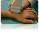
The ultrasound of the musculoskeletal has changed in recent years the daily clinical practice in rheumatology, with multiple and useful applications. Studies over a number of years have shown that it is more sensitive than the clinical examination for the detection of arthritis, tendinitis, enthesopathy, and much more reliability than simple x-ray imaging for bone erosion in inflammatory diseases. In addition, drug infusions made under ultrasound guidance seem to have better results than classic "blind" tweaks. Particularly important advantages over other imaging techniques are the low cost, the possibility of immediate application during the clinical examination and the clinical correlation of the imaging findings when the examiner is the treating physician.
|
Apps
|
>> Inflammatory arthritis |

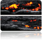
The need for early diagnosis of arthritis, when still at an early stage, for determining the severity of the disease, and for closely monitoring the response to treatment, has made ultrasound in recent years a particularly useful application to inflammatory arthritis. The visualization of a) collecting fluid in the joints, b) synovitis, inflammation of the synovial membrane surrounding the joint, c) erosions, and d) escorts of arthritis of findings (tendoneluritis, folliculitis etc) are achieved with ultrasound with high precision and sensitivity. The power Doppler signal (increased blood flow imaging) can also be used to monitor the response to treatment.
|
|
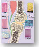
Thinning of the articular cartilage (crunchy) that overlaps the hinge and leads to the narrowing of the medial space (the distance between the two bones that make up the joint), the presence of fluid around and behind the joint (Baker's knee) Osteophytes in small and large joints and, in particular, the absence of significant synovitis (inflammation of the synovium), are some of the typical findings that can be accurately depicted by ultrasound and determine the type of treatment.
|
|

Over the last decade, ultrasound has become the method of choice for examining tendons. Tendonellitis (inflammation of the tendon cavity), tendonopathy (inflammation of the tendon itself) and rupture of the tendons can be examined with ultrasound, as it appears to be comparable to MRI, while offering the additional advantage Dynamic examination (examination during tendon movement). This is particularly useful for the emergence of periarthritis of the shoulder, as well as targeted corticosteroid infiltration.
|
|
Ultrasound has a high sensitivity in the detection of intrathetis, inflammation, ie, where the tendons, ligaments, etc. are buried on the bones. Overlapping insertions, epicondylitis, erosions, interstitials and accompanying folliculitis are some of the usual ultrasound findings.
|
|
The most common nerve trapping syndrome is carpal tunnel syndrome where ultrasound can easily locate the middle nerve and the area where pressure is exerted, while at the same time it can provide information about the cause of the pressure (in the presence of tinnitus, ganglion, Etc) and to guide targeted infiltration of the surrounding area with cortisone.
|
|
>> Crystallized arthritis |
Both urinary and pseudo-arthritic arthritis are characterized by particular ultrasound findings, which can help distinguish them from other inflammatory arthritis.
|
|

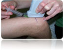
Aspiration of synovial fluid, therapeutic administration of substances intra-articular (in the joint), nerve and soft tissue infiltration, as well as biopsies of various lesions, have so far been blind. Ultrasound can be used to guide needle placement to facilitate any interventional medical practice in rheumatology such as fluid suction, bladder decompression, biopsies, local drug injections and nerve filtration, elevating the success rate to 96% and significantly reducing injuries to Adjacent molecules, pain and duration of the intervention procedure. Particularly useful is the above application of ultrasounds when the joints or fluid collections are deep and are not palpable or when they are close to anatomical structures that could be severely damaged by the puncture.
|
|
Conclusions
Although no imaging technique alone can replace the clinical examination and the detailed history, there is now plenty of data on the usefulness of musculoskeletal ultrasound in a large number of clinical applications in rheumatology. Its contribution to early diagnosis, close monitoring, and precise and safe guidance of rheumatological diseases are seen to be significant and promising. The training of rheumatologists in ultrasound performance seems to be constantly gaining ground, and the latest generation of machines promise even better performance and more applications. Thus, in the coming years, ultrasound will be a useful and indispensable tool for the comprehensive examination of patients with rheumatic and other musculoskeletal disorders.

Active view
Arthritis and arthritis
Tonsillitis
Using Power Doppler.
The use of ultrasound in Rheumatology
A new and important application
Nearchus 18 | Chania
Telephone: 2821008940
Mobile: 6973356790


